-
 在线咨询
在线咨询

关注微信公众号
课题案例系列之:
外泌体(Exosomes)
案例简介>>
 外泌体(exosomes)是直径在100nm左右,来源于内体(endosome)细胞质膜内陷形成的多泡体,多泡体与其他细胞内囊泡和细胞器融合,为外泌体的形成提供了不同类型的原料。
 外泌体作为细胞间通讯的工具, 在体液中游走, 到达靶点后, 可以 作用于另一种细胞, 引发受体细胞产生应答。外泌体对受体细胞作用目 前已有很多相关研究。细胞吸收和分泌外泌体的过程彼此相联系,外泌 体不同的吸收途径和机制以及对不同类型细胞作用的特异性增加了外泌 体在细胞间通讯功能的复杂性。外泌体在发育, 免疫应答,疾病发生, 组织器官修复等生物学过程中均发挥着重要作用。
技术路线>>
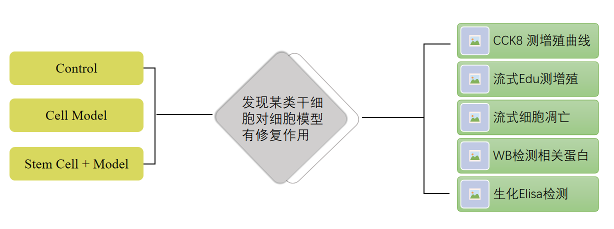
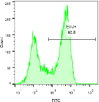
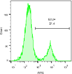
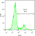
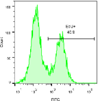
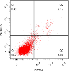
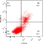
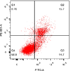
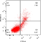
结果展示>> Fig1
A B

C D
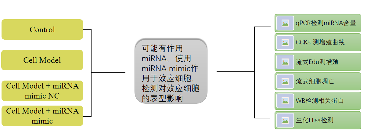
A. The CCK8 experiment verified that after co culturing stem cells with model group cells, the inhibition of cell proliferation in the model group was alleviated;
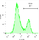
B. Edu flow cytometry detection verified that after co culture of stem cells with model group cells, the proliferation activity of model group cells was restored;
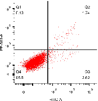
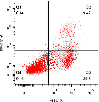
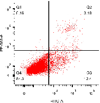
C. The flow cytometry apoptosis experiment verified that after co culturing stem cells with model group cells, the apoptosis rate of the model group cells was alleviated;
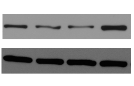
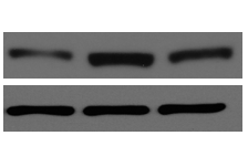
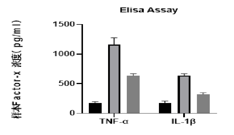
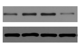
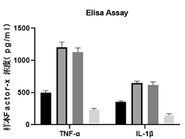
D. WB and ELISA experiments verified that after co culture of stem cells with model group cells, there were changes in cell related apoptosis and inflammatory indicators in the model group;
技术路线>>
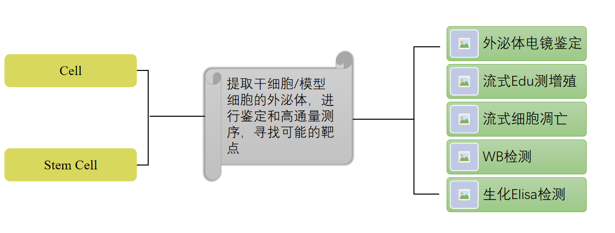
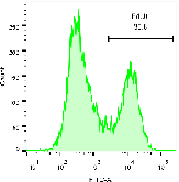
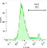
结果展示>> Fig2
A B
C D E
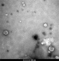
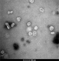
A. Transmission electron microscopy experiments were conducted to determine whether the separated extracellular vesicles have a standard bilayer membrane structure;
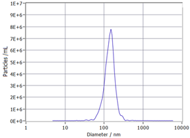
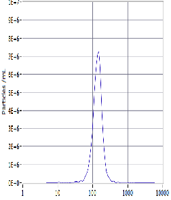
B. The particle size distribution of extracellular vesicles obtained from NTA experimental detection;
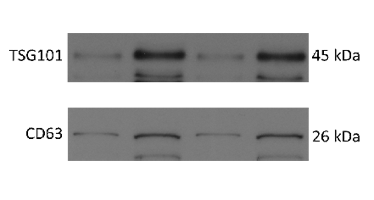
C. WB experiment detects the expression of extracellular vesicle related markers;
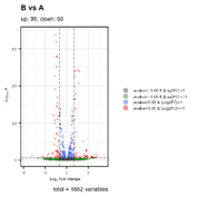
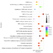
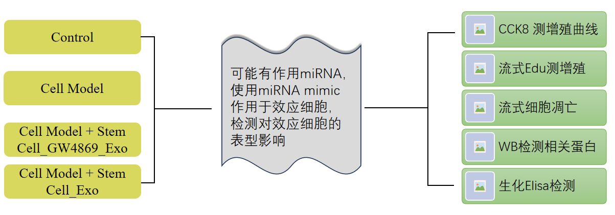
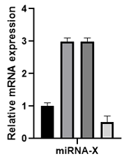
D. High throughput miRNA sequencing experiments detect differential miRNAs in samples and perform pathway analysis to screen out the most likely miRNA targets;
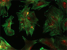
E. The exosomes are labeled with PKH26, and the cells are stained with ghost pen cyclic peptide. Exosomes tracing technology shows the phagocytosis of the exosomes.
技术路线>>
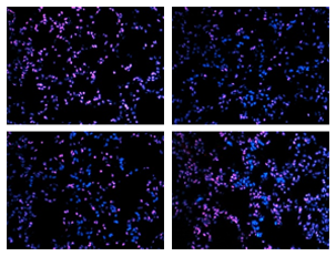
结果展示>> Fig3
A B C


D E
技术路线>>
结果展示>> Fig4
A B C
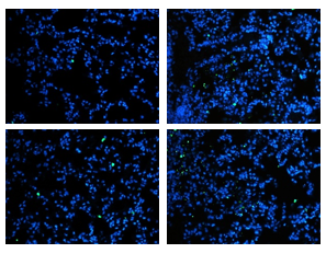
D